

TIENDA
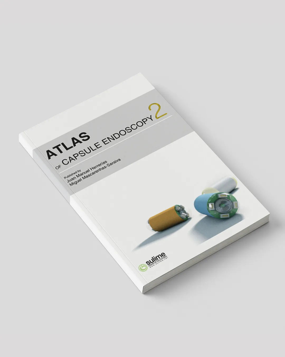
Atlas of capsule endoscopy 2
It is a great privilege and a real pleasure for me to have been invited to write the foreword for the 2nd edition of Atlas of Capsule endoscopy published by my prestigious friends Juan Manuel Herrerías and Miguel Mascarenhas-Saraiva. It has been my good fortune to have known these two distinguished physicians for many years and to have seen them grow as respected clinicians and educators in the field of gastroenterology.
Until a few years ago, the small bowel was an organ which was very difficult to explore with the available endoscopic, radiological and nuclear medicine techniques. In routine practice only the last few centimeters of the ileum were accessible to retrograde visualization by ileocolonoscopy. Explorations from the proximal side by push, sonde or intraoperative enteroscopy were invasive procedures that do not always allow us to visualize the lesions in the small bowel. Sonde enteroscopy had been abandoned in the 90's because it was a tedious exploration (long duration of the procedure) and it had several technical limitations. Push enteroscopy is limited by the depth of insertion of the scope and it is poorly tolerated. Intraoperative enteroscopy is the most effective of these techniques, but is the most invasive with an important percentage of adverse side effects.
I witnessed Dr. Paul Swain first presenting the use of the wireless capsule endoscope in May 2000 at Digestive Disease Week in San Diego, during the Plenary Session of the American Society for Gastrointestinal Endoscopy, there was a tremendously enthusiastic response. Comparisons were made to the miniaturized spaceship used to examine the body's inner spaces in the science fiction movie "Fantastic Voyage".
Capsule endoscopy was launched at the beginning of this millennium and since then has had a very important impact on managing obscure gastrointestinal bleeding and many other small bowel diseases. The initial capsule endoscope was developed by Given Imaging (Yoqneam, Israel) and approved in Europe by the European Agency and in the United States by the Food and Drug Administration in 2001.
With Wireless capsule endoscopy (CE) we can provide a simple, safe, non invasive, reliable, procedure, well accepted and tolerated by the patient, which has revoluzioned the study of the small bowel. This technique evaluates endoscopically, with high resolution images, what has been called "the last frontier" of endoscopy, the small bowel, avoiding any sedation, surgery or radiation exposure.
Currently CE is recommended as a third stage examination, after negative gastroscopy and colonoscopy in patients with obscure gastrointestinal bleeding. Also many studies have established, with a growing body of evidence, that this technique is cost-effective in other clinical situations, such as detection of small bowel lesions in Crohn's disease in patients in which other methods fail to prove the diagnosis, non steroidal anti-inflammatory drug enteropaties, celiac disease, small bowel polyposis syndromes and small bowel tumors. Other possible indications are HIV patients with gastrointestinal symptoms, malabsortive syndromes other than celiac disease, Henoch-Schönlein purpura, patients with small bowel transplants and with intestinal graft versus host disease, particularly in monitoring the response to immunosuppressive therapy.
The acquired knowledge of the wide range of lesions that can be found in the small bowel, encouraged the implementation of some diagnostic and therapeutic techniques, such as double balloon enteroscopy, MRI-enteroclysis and CT-enteroclysis.
The main contraindication of performing the CE is the suspicion or knowledge of an obstruction in the GI tract.
The device retention is the main complication of the procedure and is defined when CE remains in the digestive tract for a minimum of 2 weeks. The frequency of this problem varies, depending mostly on the clinical indication for CE, and ranges from 0% in healthy subjects, to 1.5 %in patients with obscure GI bleeding, to 5% in patients with suspected Crohn's disease and to 21% in patients with intestinal obstruction.
At present CE has some technical limitations, it can not be used to obtain biopsy specimens or for endoscopic treatment and it can not be controlled remotely. CE has also some clinical limitations which are the problem in sizing and locating small bowel lesions, a possible false-negative CE result, due to the fact that the global miss rate is about 11%, ranging from 0.5% for ulcerative lesions to 18.9% for neoplastic disease and the fact that some times we can get findings of uncertain relevance in healthy subjects. Other drawback is that in almost 20% of procedures the capsule does not reach the cecum while it is active.
This technique is available in over 5000 gastrointestinal centers throughout the world.
Since its arrival, more than 650,000 capsules have been swallowed worldwide and more than 1000 peer-reviewed publications have appeared in medical literature. The most important Gl societies have published guidelines about its use.
In latter years, breakthrough developments in CE technology have enabled the direct visualization of the upper and lower segments of the gut using specifically designed capsules.
This updated second edition of the atlas is much improved compared to the first edition; many new chapters, authors and technological advances have been added.
The editors have chosen the authors of each chapter very well, from eight different countries, with a mixture of established leaders and rising younger colleagues who represent the next generation staking its claim to this rapidly evolving field of the gastrointestinal endoscopy.
The images are well chosen most of them of high quality and superbly produced, raising the exceptionally high quality of the first edition.
The atlas is divided into six parts. The first part consists of 10 chapters and covers general aspects of the technique. The second part deals with its usefulness for the study of the esophagus, the third shows the possibilities that the capsule gastroscopy presently offers as well as the findings that we can see in the stomach when we are performing an exploration with the capsule. The fourth consists of 16 chapters and deals with the multiple applications that this technique has in the small intestine, including motility studies, and with the alternative techniques for enteroscopy. The fifth part deals with capsule colonoscopy and the possibility of performing pan-endoscopy with the colon capsule, the sixth part discusses the utility of capsule endoscopy in pediatric patients, in patients with abdominal pain and finally the future developments of capsule endoscopy.
I believe that this atlas has much to offer to individuals at all levels of involvement in the field of gastroenterology, from students to even the most seasoned clinicians. And finally, I want to congratulate not only the publishers but also the authors for their excellent contributions to this atlas.
Miguel Muñoz-Navas MD, PhD
Director Gastroenterology Department
University of Navarra Clinic
School of Medicine. University of Navarra. Pamplona. Spain.