

TIENDA
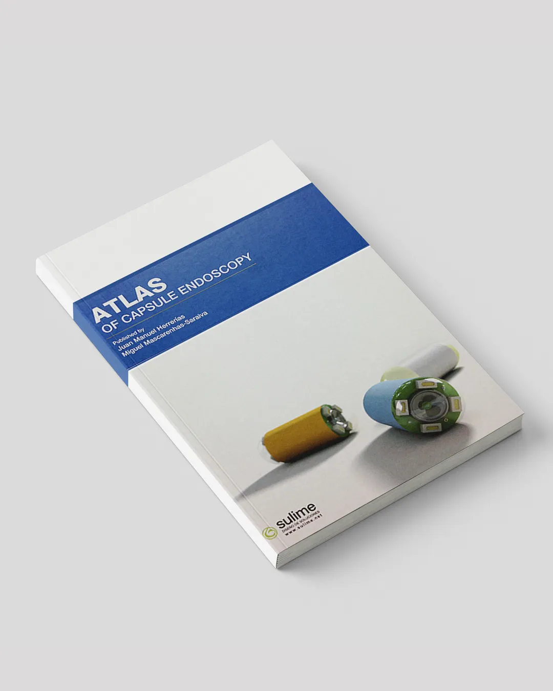
Atlas of capsule endoscopy
Since the appearance of the first known "endoscope", designed by Phillip Bozzini around 1806, and the somewhat more helpful instruments devised a little later by Fisher, Ségalas and Désormeaux, the quest of the curious physicians to make the innermost parts of the body visible by the human eye seems to know no bounds. In 1853 Désormeaux succeeded in having his instrument manufactured and sold commercially by the firm Charrière. By the end of the 19th century, rigid endoscopes with lighting systems in their shaft had been designed and were employed until the mid-20th century. They were then replaced by flexible instruments taking advantage of Van Heel, Hopkins and Kapany's discovery of optical fibers.
Their practical application was the work of Basil Hirschowitz and Larry Curtiss, and the fiberscopes which emerged in the late 1960s seemed to be the lasting development achievement in the visualization of the gastrointestinal tract as photographs of reasonable definition could be taken and biopsy samples could be collected. However, technology continued its unstoppable march towards further achievements.
During the combined meeting on the American Gastroenterological Association (A.G.A.) and the American Society of Gastrointestinal Endoscopy (A.S.G.E.) in 1983, the firm Welch-Allyn, mostly known for manufacturing ophthalmologic equipment, exhibited a new instrument that caused tremendous impact: the video-endoscope. It looked just like an ordinary fiberscope except for the objective lens which had been replaced by a small CCD sensor of only a few millimeters in diameter. Many of us stared in awe at the images transmitted with excellent definition onto a television screen. The "chip" revolutionized medical and surgical endoscopy and rapidly began to supplant the previous models. With these new videoscopes that could be manufactured with very small diameters, it seemed we had reached the desideratum in endoscopic visualization, where any type of endoscopy, such as bronchoscopy or cystoscopy, could be performed with little discomfort for the patient. Pediatric gastrointestinal endoscopy became equally easy.
About a decade ago, however, possibly inspired by the radiotelemetering capsules that Jacobson had devised at the Karolinska Institute for measuring and recording temperatures, pH and pressures within the gut, some eager scientists started experimenting with very small CCD sensors devised which were able to transmit images from the gut to an outside monitor. Various types of capsules in which a sensor was enclosed could be easily swallowed and the resulting images could be followed throughout the gut.
Two separate teams of researchers working independently at different locations succeeded in transmitting images of the lumen of the gut using sensors enclosed in rather small pills. These initial images were already of reasonable quality. An Israeli team led by a physicist, Gavriel Iddan, designed the first wireless endoscopy capsule. Paul Swain, a surgeon from the London Hospital, who had joined up with them, was the first to test a prototype on himself. Many of us watched him in the act of swallowing the capsule in public or saw this event later on a video that was shown at several meetings. The outcome was the successful production of a commercially available video capsule which was soon tested in many academic hospitals with apparent success. It was not long before gastroenterologists realized that the new device could detect the cause of obscure cases of gastrointestinal bleeding, especially those originating from lesions in the small bowel, an area not visualized by ordinary fiberoptic instruments. Angiectasias, submucosal tumors and superficial ulcerations, such as those occurring in early inflammatory bowel disease, were successfully identified.
This excellent atlas is the work of two renowned specialists and their teams, Miguel Mascarenhas-Saraiva, son of a distinguished surgeon from Porto, and Juan M. Herrerías from the University of Sevilla. Several of their colleagues with wide experience in the field have contributed chapters: they include the developers of the capsule, Gavriel Meron, Ellen Fischl and Eitan Scapa from Israel, Michel Delvaux and Gerard Gay from Nancy University, Marco Pennazio from the University of Torino as well as Miguel Muñoz-Navas and his colleagues from Navarra University.
The book is divided into three parts. Part I deals first with the birth and development of capsule endoscopy, written by the Israeli team. It is followed by Mascarenhas Saraiva's description of the procedure and the indications for capsule endoscopy, and an assessment of the contraindications and complications of the procedure by Pennazio. Next comes important new terminology of capsule endoscopy, adapted by Delvaux from the minimal terminology standards set up by the European Society of Gastrointestinal Endoscopy and other organizations. Finally, normal anatomy as seen through the capsule is described in detail by the Sevilla team.
Part II deals with diseases of the small bowel; iatrogenic lesions, neoplastic and vascular disorders, malabsorption syndromes, surveillance of polyposis and the use of the capsule in pediatric gastroenterology are very well documented.
Part III is dedicated to other uses of the capsule. There are two chapters by the Seville group on the new esophageal capsule, including a description of the procedure and their findings in several esophageal conditions. A useful chapter on the so-called "patency" capsule follows. This device should be swallowed before the diagnostic procedure and will disintegrate almost completely above a stenotic area of the gut, thus avoiding the danger of blockage with an ordinary diagnostic capsule.
The book ends with a chapter on the recently devised colonic CamPill by Muñoz-Navas and his team from the University of Navarra. They show that pill-colonoscopy is a feasible and safe procedure which in most cases allows screening of the entire colon.
Dictionaries describe an atlas as a "bound collection of plates illustrating any subject". As such, the quality of the illustrations is of paramount importance in a book of this kind. The authors have succeeded in gathering a series of images of excellent definition from all parts of the gut. Particularly impressive are those of small flat ulcerations and aphthous lesions, vascular ectasias and small tumors which went unrecognized until now. There are many pictures of the different types of capsules as well as clear algorithmic depictions of various decision-making situations. The authors show that we can confidently predict that capsule endoscopy will replace most of the current endoscopes for screening, diagnosis and follow-up purposes in the near future.
Miguel Mascarenhas-Saraiva and Juan Manuel Herrerías are to be heartily congratulated for producing such a valuable book. It should be required reading and readily available for all those interested in gastrointestinal endoscopy, including medical and surgical gastroenterologists and general internists.
Francisco Vilardell MD DSc FRCP FRCP (Ed) FACP
Director Emeritus, Postgraduate School of Gastroenterology,
Autonomous University, Hospital Sant Pau, Barcelona.